St Ives Physiotherapy & Sports Therapy Centre
  • Home
  • About
    • Our Team
    • Careers
    • Privacy Policy
  • Services
    • Studio Timetable
    • Who We Treat
    • Conditions Treated
    • Methods of Treatment >
      • Dry Needling
    • Consultation Information
    • CET Classes
    • Back Classes
    • Youth Strengthening
    • Tai Chi
    • Village Walkers
  • Telehealth
  • Products
    • Exercise Products
    • Massage and Foam Rollers
    • Braces and Support
    • Tapes and Bandages
    • Hot and Cold Therapy
    • Orthotics and Footcare
    • Books and DVDs
  • Resources
    • COVID-19
    • Exercise Videos
    • Blog
    • Informative Videos
    • Newsletters & Brochures
    • Sports Affiliations
    • Related Websites
  • Contact Us
    • Feedback

PLANTAR FASCIOPATHY             (formerly called 'Plantar Fasciitis')

27/3/2017

0 Comments

 
Picture

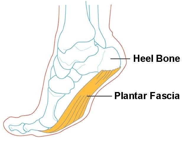
Plantar fasciopathy (also known as plantar fasciitis) is an overuse condition of the plantar fascia at its attachment to the calcaneus (heel bone).  It is the most common cause of plantar heel pain, which has an estimated prevalence of 4-7% of the population.
 
Anatomy
The plantar fascia is a strong band of tissue (like a ligament) that stretches from your heel bone to your middle foot bones.  It supports the arch of your foot and also acts as a shock absorber in your foot.
 
Common Causes/Risk Factors
Plantar fasciopathy commonly affects very physically active people (e.g. runners) or people with high amounts of standing occupations work (who may also have a high BMI).
 
Only one true prospective study has investigated risk factors for plantar fasciopathy.  The study was conducted in a cohort of 166 runners followed over 5 years.  The study found the overall incidence of plantar fasciopathy over the 5 year period to be 31%. 
 
Four risk factors that were significantly associated with an increased risk of plantar fasciopathy were:
  1. Varus knee alignment (knee collapsing inwards)
  2. Use of spiked athletic shoes
  3. Cavus foot posture (rigid, high arch)
  4. Greater number of practise days per week
 
Other risk factors for plantar fasciopathy include:
  • If you are on your feet for a lot of the time, or if you do lots of walking, running, standing, etc, when you are not used to it or have previously had a more sedentary lifestyle.
  • If you have recently started exercising on a different surface - for example, running on the road instead of a track.
  • If you have been wearing shoes with poor cushioning or poor arch support.
  • If you are overweight - this will put extra strain on your heel.
  • If there is overuse or sudden stretching of your sole. For example - athletes who increase running intensity or distance; poor technique starting 'off the blocks', etc.
  • If you have a tight Achilles tendon (the big tendon at the bottom of your calf muscles above your heel). This can affect your ability to flex your ankle and make you more likely to damage your plantar fascia.
 
Signs and symptoms
Pain is usually felt on the underside of the heel, and is often most intense with the first steps of the day or after rest.  It is commonly associated with long periods of weight bearing or sudden changes in weight bearing or activity.
You may experience:
  • Sharp pain in the inside part of the bottom of the heel.
  • Heel pain that tends to be worse with the first few steps after awakening, when climbing stairs or when standing on tiptoe.
  • Heel pain after long periods of standing or after getting up from a seated position.
  • Heel pain after, but not usually during, exercise.
  • Mild swelling in the heel.
 
What about Scans or X-rays?
If a patient has the classic history and examination findings and there is no other suggestion of a ‘red flag’ then medical imaging or other investigations are not indicated.
 
Further investigation is recommended if:
  • Pain is particularly persistent without responding to treatment after greater than one month
  • If the nature of the pain is atypical for plantar fasciopathy (e.g. acute onset of pain, persistent pain even at rest, systemic symptoms, night pain).
 
Blood tests may be warranted to rule out inflammatory arthritis, which can cause symptoms in the heels, although frequently symptoms will be more widespread.  The primary imaging modalities that are useful include plain radiographs (X-rays), ultrasound or MRI.
 
What about a Heel Spur?
X-rays may sometimes show a heel spur; however, heel spurs are also common in people without heel pain, and the presence of a spur does not indicate that this is the cause of the pain.  X-rays of both feet are useful as they may show spurs bilaterally and allow comparison of the feet in unilateral symptoms.
 
Treatment
Treatment is often multi-faceted and generally starts with conservative interventions, modifying load and addressing any risk-factors.  A physiotherapist will usually assess your biomechanics and look at all the links in the chain, from the foot right up to the hip, pelvis and core.
 
Some authors define treatment into First, Second and Third-Tier treatment which essentially relate to short-, medium- and long-term options.  The tiers also relate to the complexity, cost and invasiveness of the intervention.
First Tier/Short Term Treatment
Aims: minimise the aggravating activity and educate the patient about the potential causes of their heel pain.  Discuss and modify training loads as appropriate.
 
  • Modify footwear as appropriate, consider sue of soft silicone gel heel pads or contoured orthotics.
  • Decrease or avoid activities that cause the heel pain to worsen.
  • Discuss potential options to decrease the hours of standing per day if that is likely a contributing factor.
  • Taping
  • Strengthening exercises
  • Stretches
  • Massage
  • Weight-loss if appropriate
 
Second Tier/Medium Term
  • Dry needling
  • Foot orthoses
  • Corticosteroid injection
  • High-load strength training
 
Third Tier/Long Term
  • Night splints should be considered in patients with pain of over 6 months in duration
  • Extracorporeal shockwave therapy may be of benefit for longstanding cases
  • As the evidence for surgery is unclear and of low quality, it should only be considered in severe longstanding cases that do not respond to any of the evidence-based treatments available.
 
 
REFERENCES:
Brukner P, Khan K, et al.  Clinical Sports Medicine.  Volume 1, Injuries.  5th Edition. 2017.  McGraw Hill Education.
 
http://sma.org.au/resources-advice/injury-fact-sheets/plantar-fasciitis/  Plantar Fasciitis Injury Fact Sheet, Sports Medicine Australia.
 
 
0 Comments

Your comment will be posted after it is approved.


Leave a Reply.

    Author

    David McWilliams graduated from Macquarie University with a Doctor of Physiotherapy. He is a member of Australian Physiotherapy Association, Sports and Exercise Physiotherapy Australia, Sports Medicine Australia

    Archives

    April 2019
    May 2018
    March 2017
    December 2016
    October 2016
    August 2015
    July 2015
    June 2015
    April 2015

    Categories

    All

St Ives Physiotherapy & Sports Therapy Centre © 2020
All Rights Reserved
  • Home
  • About
    • Our Team
    • Careers
    • Privacy Policy
  • Services
    • Studio Timetable
    • Who We Treat
    • Conditions Treated
    • Methods of Treatment >
      • Dry Needling
    • Consultation Information
    • CET Classes
    • Back Classes
    • Youth Strengthening
    • Tai Chi
    • Village Walkers
  • Telehealth
  • Products
    • Exercise Products
    • Massage and Foam Rollers
    • Braces and Support
    • Tapes and Bandages
    • Hot and Cold Therapy
    • Orthotics and Footcare
    • Books and DVDs
  • Resources
    • COVID-19
    • Exercise Videos
    • Blog
    • Informative Videos
    • Newsletters & Brochures
    • Sports Affiliations
    • Related Websites
  • Contact Us
    • Feedback